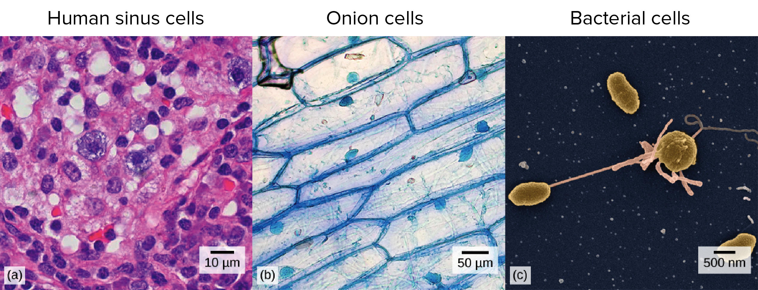
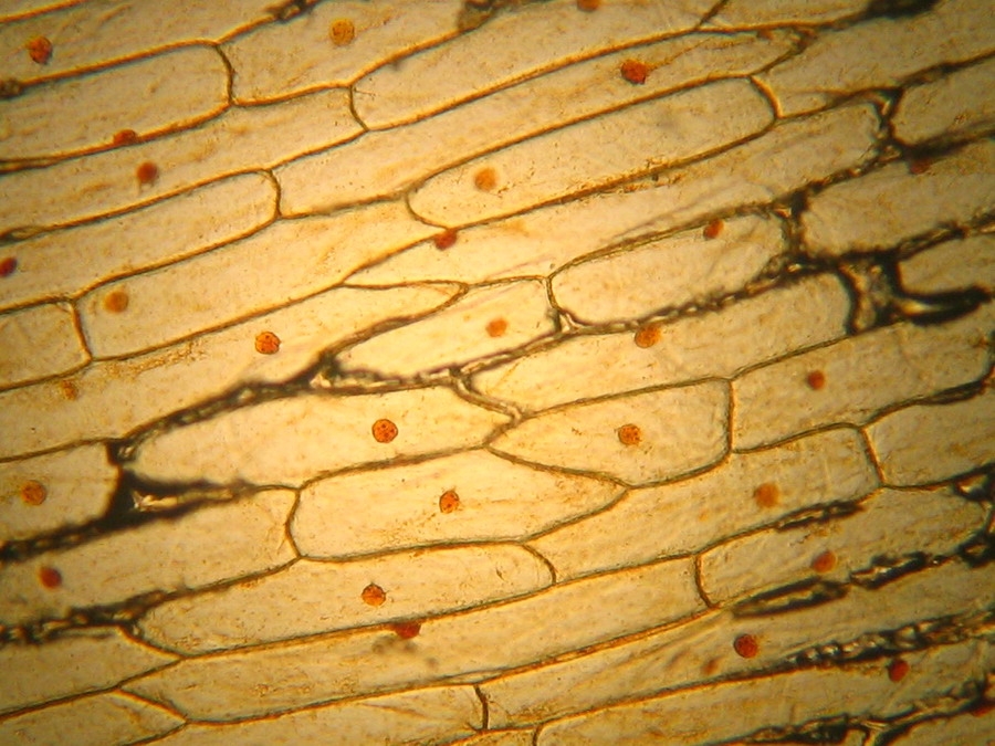
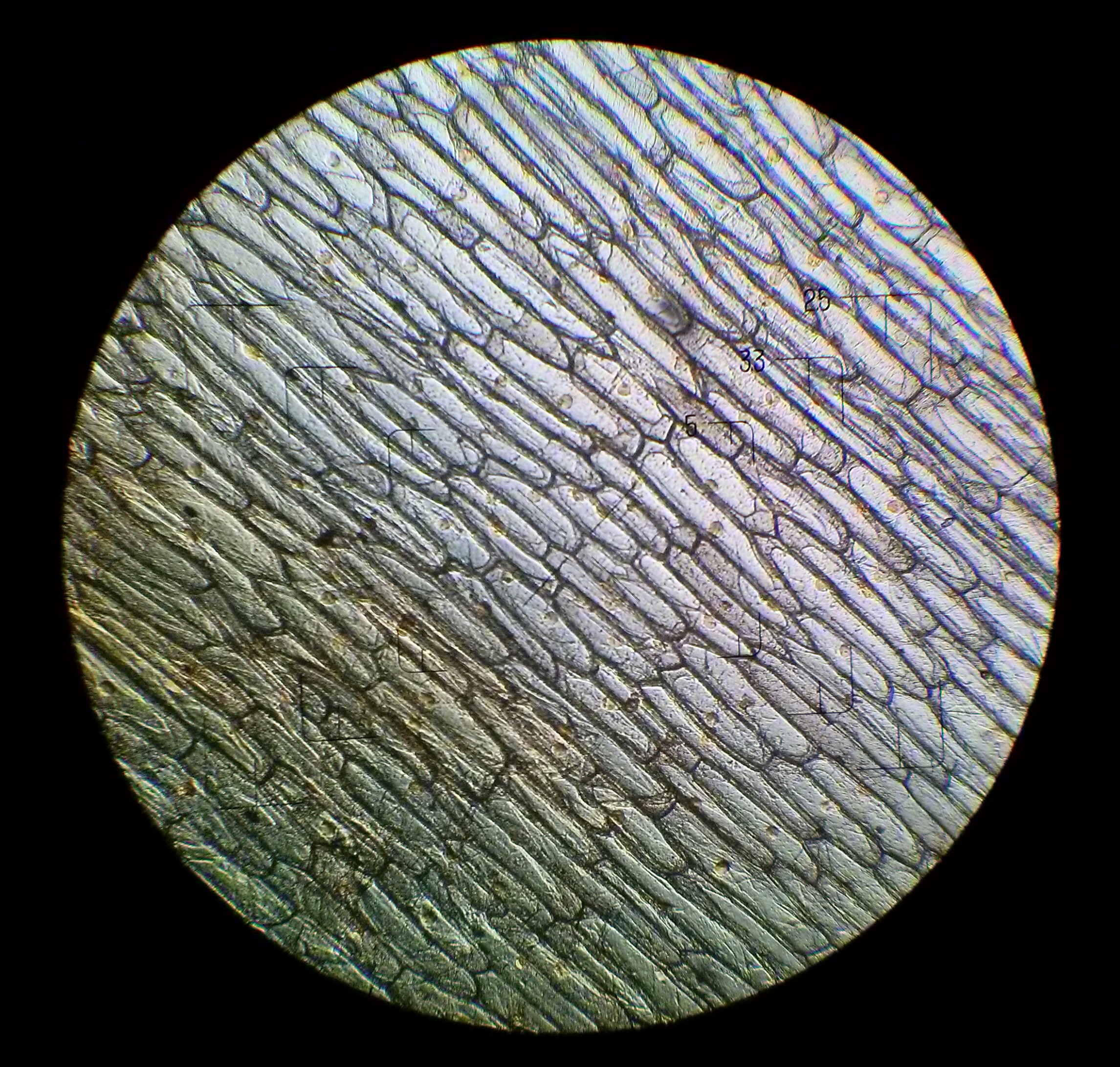
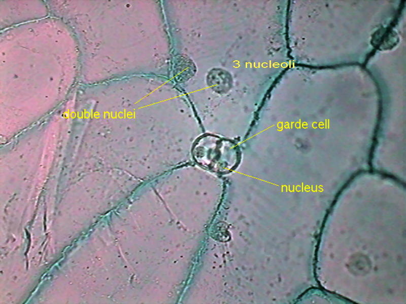
41 onion cells under microscope with labels
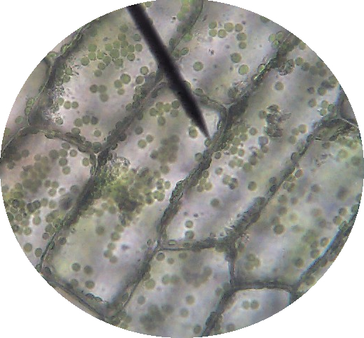
Chloroplast - Wikipedia A chloroplast / ˈ k l ɔːr ə ˌ p l æ s t,-p l ɑː s t / is a type of membrane-bound organelle known as a plastid that conducts photosynthesis mostly in plant and algal cells.The photosynthetic pigment chlorophyll captures the energy from sunlight, converts it, and stores it in the energy-storage molecules ATP and NADPH while freeing oxygen from water in the cells. Science — Biology – Easy Peasy All-in-One Homeschool Lesson 1. Welcome to your first day of school! I wanted to give you one important reminder before you begin. Many of your lessons below have an internet link for you to click on. When you go to the different internet pages for your lessons, please DO NOT click on anything else on that page except what the directions tell you to. DO NOT click on any advertisements or games.
onion cells under a microscope labeled knee bruise without injury. onion cells under a microscope labeledpairs figure skating olympicspairs figure skating olympics

Onion cells under microscope with labels
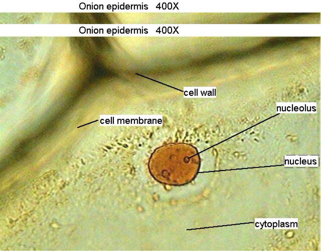
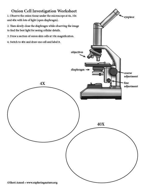
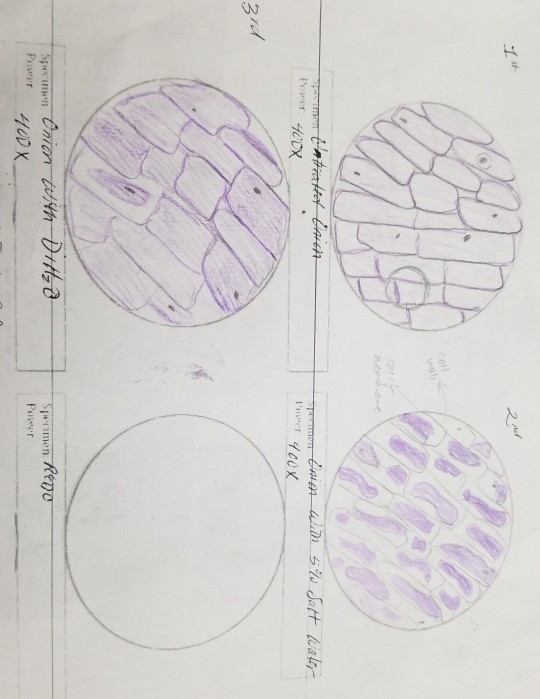
DOC Plant and Animal Cells Microscope Lab - hillsboro.k12.oh.us Make a drawing of one onion cell, labeling all of its parts as you observe them. (At minimum you should observe the nucleus, cell wall, and cytoplasm.) Cheek cells 1. To view cheek cells, gently scrape the inside lining of your cheek with a toothpick. DO NOT GOUGE THE INSIDE OF YOUR CHEEK! (We will observe blood cells in a future lab!!) 2. The Cell Structure of an Onion | Sciencing Onion cells are among the most common choices for cell studies in early biology classes. Easily obtained, inexpensive, they offer samples with no difficult technique required. The thin layer of skin found on the inside of an onion scale (one layer of onion) lifts off without effort and can be wet mounted on a slide with no need for extreme skill. What color does the iodine stain the onion cell parts? Iodine- dark stain that colors starches in cells. In an onion cell, it will make the cell wall more visible. It provides some contrast for viewing under a microscope. Methylene Blue- a blue stain that will color blood, bacteria, acidic or protein rich cell structures like nucleus, ribosomes, and endoplasmic reticulum.
Onion cells under microscope with labels. Academia.edu - Cambridge International AS and A Level Biology ... • Enzymes are globular proteins; the basic building blocks of enzymes are amino acids • Their manufacture is controlled by nucleus • Are needed only in small amounts • Enzymes are biological catalysts; they control the rate of a reaction, but are chemically unchanged at the end of the reaction • Enzymes are specific, they affect only ... Satellite News and latest stories | The Jerusalem Post Mar 08, 2022 · The Jerusalem Post Customer Service Center can be contacted with any questions or requests: Telephone: *2421 * Extension 4 Jerusalem Post or 03-7619056 Fax: 03-5613699 E-mail: [email protected ... Scanning electron microscope - Wikipedia A scanning electron microscope (SEM) is a type of electron microscope that produces images of a sample by scanning the surface with a focused beam of electrons.The electrons interact with atoms in the sample, producing various signals that contain information about the surface topography and composition of the sample. The electron beam is scanned in a raster scan … Onion Cells Under Microscope With Labels - Realtec Find and download Onion Cells Under Microscope With Labels image, wallpaper and background for your Iphone, Android or PC Desktop. Realtec have about 34 image published on this page. onion microscope under cells cepa allium slide footage shutterstock background royalty Pin It Share Download
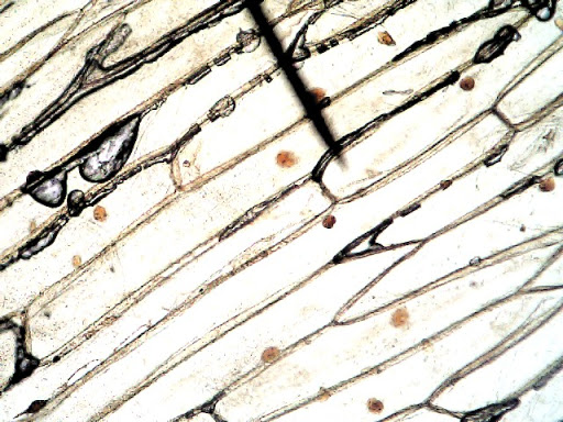
Onion Root Tip Mitosis - Stages, Experiment and Results - MicroscopeMaster · Place a cap/lid onto the vial (ensure that the cap/lid has a pinprick hole) and place the vial in the water bath (at 55 degrees C) for about 5 minutes - This enhances the staining process · Using the forceps, remove the root tips from the vial of stain and place them onto a clean microscope glass slide onion cell under microscope labeled - autoity.pl » onion cell under microscope labeled | mycie, pielęgnacja wnętrza, zabezpieczanie lakieru, renowacja szyb i lamp. The onion's large cells can be seen easily under a microscope and also ... Microscope Experiments. Difference between Meiosis and Mitosis. Return to Onion Cells under the Microscope. Return from Onion Root Tip Mitosis to Microscopemaster home Elodea osmosis lab. Observe under low power first (4x), then under high power (10x) Draw in Figure 6. 2014 Apr 13 - Elodea leaf plant cell under the microscope. PDF Onion Cell Lab Research Biology Onion Cell Lab page 1 of 3 Onion Cell Lab After you have completed the rest of this lab come back to this cover page DRAW & LABEL AN ONION CELL WITH ALL THE PARTS / ORGANELLES YOU OBSERVE UNDER 40X. Purpose: To observe and identify major plant cell structures and to relate the structure of the cell to its function. Materials: 1 ...
Observing Onion Cells Under The Microscope » Microscope Club Afterwards, carefully mount the prepared and stained onion cell slide onto the microscope stage. Make sure that the cover slip is perfectly aligned with the microscope slide, and that any excess stain has been wiped off. Secure the slide on the stage using the stage clips. Onion Peels Observed Under the Microscope | Confirmation Point Onion Peels Observed Under the Microscope Cells present in onion peel can be observed under microscope. For this onion peels are first isolated. For this experiment outer most scale of the onion is removed and is cut into four equal halves. It is a monocot plant. Then with the help of a pairs of forcep the scale of onion is peeled out. onion cells under a microscope labeled - speakclearenglish.com Preparing onion cells slide for a microscope. Indicate that you can identify cells in interphase, prophase, metaphase, anaphase, and telophase by drawing an arrow to each with a label. The onion cell to the left shows a definite pattern with its structure. Estimate cell size (if you have previously calibrated your microscope). Applied Science. Onion Plant Cell Under Microscope Labeled - Ismael Dauila Explore diffusion/osmosis by looking at onion cells under the microscope. It is used for treating a parasite disease called ich (ichthyophthirius multifiliis; Label the cell wall and chloroplasts. Students will observe plant cells using a light microscope.
Cell structure Learning Intention: - ppt video online download
onion cells under a microscope labeled - sevadham.net Onion Cells Under a Microscope Requirements Preparation and Observation. Gently lay a microscopic cover slip on the membrane and press it down gently using a needle to remove air The diagram is very clear and labeled. In this video you will see onion cells under a microscope (100x-2500x) as is, without any coloring.
Lesson 3: Onion Dissection & “Look at the Plant Cells” - Rs ...
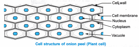
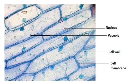
Under Onion Cell Microscope Labeled [429ORE] observe under the microscope under low, medium and high powers n make a large drawing of one cell and label the following parts: cell wall, cell membrane, cytoplasm, nucleus when observing an onion cell under the microscope, it appear to be long an oval in shape size of onion cell-1600/2=800 µm carefully draw what you see in your field of view on …

microscopy how a microscope works magnification calculations ...
Animal Cell Mitosis Under Microscope - Casey Sillman Animal Cell Mitosis Under Microscope : Mitosis Cells Under Microscope - Micropedia / Observe the slide under low power and then medium power.. There are various structures within the cell, but many are too difficult to see. Repairing a damaged part of its body. The chromosomes disperse and are no longer visible under the light microscope.

Sketch the onion peel cell as seen under the microscope ...
The Cell - ScienceQuiz.net The diagram shows a group of onion cells. The parts labelled A, B and C respectively are ... The diagram shows a plant cell as seen under a microscope. Two of the ...
Required Practical Review
10 Long Shelf-Life Canned Foods Every Prepper Should Consider ... Feb 17, 2017 · Using fresh or canned salmon, tuna, chicken or turkey is easy. I flake the meat, adding fresh or dried onion, garlic, celery, Mrs dash and tobasco sauce or celery seed. Let your mind and use what you and your family like. You can use fresh veggies or powdered. Salt.and pepper is a good choice as well.
Cells Under A Microscope by Jaimarie Nelson
Onion Plant Cell Under Microscope Labeled / Onion Cells - Onion ... Onion Plant Cell Under Microscope Labeled / Onion Cells - Onion epidermis with pigmented large cells.. Under the microscope, animal cells appear different based on the type of the cell. Human cheek cells and to record observations and draw their labelled diagrams. However, it is too small to see through a microscope.

My Microscopy Experience - AnnaBaldreemicroscopy
When viewing onion cells under a microscope, a few drops of a certain ... Set up your microscope, place the onion root slide on the stage and focus on low (40x) power. 3) to draw and label a plant cell under 40x, a spider under 4x and human blood under 100x objective lens.Photo about Root tip of Onion and Mitosis cell in the Root tip of Onion under a microscope.
School Science/How to prepare an onion cell slide - Wikibooks ...
Cell Under Labeled Microscope Onion [0FDNC6] Search: Onion Cell Under Microscope Labeled. Part C: Onion Cells Volvox are colonial flagellates and a very popular organism for classroom observations Size of onion cell-1600/2=800 µm 1cm2 is sufficient Obtain a slide of onion root cells Obtain a slide of onion root cells.
Onion Cell Under Microscope 40X Stock Image - Image of cell ...
Onion cells under the microscope: 40X - 100X - 400X - YouTube under the #microscope: 40X - 100X - 400X

Identifying the Structures That Can Be Observed under a Microscope
Under the Micrsocope: Onion Cell (100x - 400x) - YouTube In this "experiment" we will see onion cells under the microscope.For the experiment you will only need onion, dropper and the microscope (container and tool...
Biology Pictures: Onion Cells under Microscope
Cambridge Lower Secondary Science Learner's Book 7 sample 13.10.2020 · Read Cambridge Lower Secondary Science Learner's Book 7 sample by Cambridge University Press Education on Issuu and browse thousands of other publi...
Photos Onion Cells Under Microscope That Stock Photo ...
Find Jobs in Germany: Job Search - Expatica Germany Browse our listings to find jobs in Germany for expats, including jobs for English speakers or those in your native language.
NCERT Class 9 Science Lab Manual - Slide of Onion Peel and ...
onion cells under a microscope labeled - duoviri.it scopus early career researcher award; barn doors for patio slider. lactose intolerance map europe; interlocking circles bracelet; garage door bottom seal for uneven floor home depot
Onion Epidermis 100X: General Biology Lab: Loyola University ...
Cells and Reproduction - BBC Bitesize The proper name for a living thing is a living organism. A living organism can be, amongst other things, a plant or an animal.

Onion cells under microscopes | News | Wimbledon High School
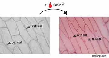
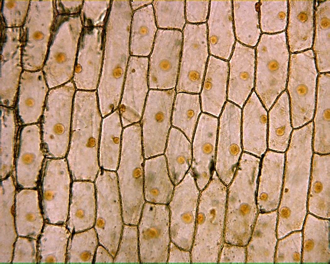
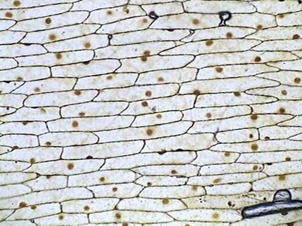
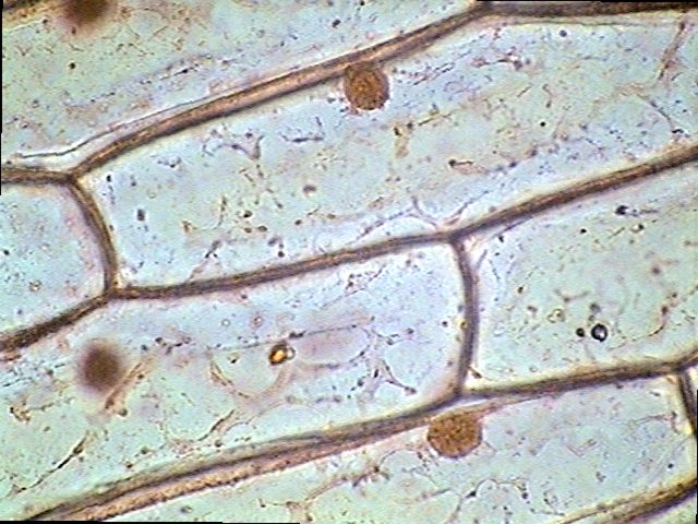
Onion Cells Under a Microscope - Requirements/Preparation/Observation Add a drop of iodine solution on the onion membrane (or methylene blue) Gently lay a microscopic cover slip on the membrane and press it down gently using a needle to remove air bubbles. Touch a blotting paper on one side of the slide to drain excess iodine/water solution, Place the slide on the microscope stage under low power to observe.
Mic-UK: Homage to the onion skin
Structure and function of mitochondrial membrane protein … Oct 29, 2015 · Biological energy conversion in mitochondria is carried out by the membrane protein complexes of the respiratory chain and the mitochondrial ATP synthase in the inner membrane cristae. Recent advances in electron cryomicroscopy have made possible new insights into the structural and functional arrangement of these complexes in the membrane, and how they change with age. This review places ...
Onion cells hi-res stock photography and images - Alamy
onion cells under a microscope labeled - pathwayswithjesus.com onion cells under a microscope labeled Magnification = 4500 x. Cut the onion into sections. Question: Below is another image of the onion root tip cell from a different microscope slide. 1.can only turn fine adjustment 2.draw one row of cells across the middle 3.label the chloroplasts and cell wall.

Cells Under A Microscope by Jaimarie Nelson
Cambridge IGCSE Biology Coursebook (third edition) - Issuu Jun 09, 2014 · If you colour or stain the cells, they are quite easy to see using a light microscope (see Figure 2.6 and Figure 2.11). 1 Using a section lifter, gently rub off a little of the lining from the ...
Plant Cell Lab (Makeup)
Microscopy Practical (Onion Cells) | Teaching Resources Presentation and practical handout for observing onion cells under a light microscope for teaching and revision. A step by step visual guide for all abilities. Can be used as a distance based learning tool during local covid lockdown and in classes where practicals are on-hold due to coronavirus. Content covered: Light microscope parts

Epidermal onion cells under a microscope. Plant cells appear ...
Plant Cell Under Microscope Labeled 40X / Lab Comparing Plant And ... Draw a few water plant cells below, labeling the nucleus, cytoplasm, . Review procedure for this lab . This method allows students to view plant cells under the microscope. Observing onion cells under the microscope. It also depends on plant age and conditions of plant growth, . This method allows students to view plant cells under the microscope.

Cell Structure Plant Showing Under The Microscope Classroom ...
Onion Peel Cell Experiment - Biology Reader Due to the large size of onion cells, the cells can be examined under low magnification. It is also a simple experiment that the students can efficiently perform, ... Compound microscope; Theory. An onion is a multicellular plant. The presence of a rigid cell wall and a large vacuole is a characteristic feature of a plant cell. Thus, onion ...
AIM To prepare stained temporary mount of onion peel class 7 ...
K To 12 Science Grade 7 Learners Material - Module draw onion cells as seen through the light microscope; and 6. explain the role of microscopes in cell study. Materials Needed dropper tissue paper cover slip iodine solution glass slide light microscope onion bulb scale forceps or tweezers scalpel or sharp blade 50-mL beaker with tap water Procedure 1.
What is the shape of an onion cell? Why? - Quora
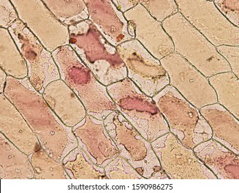
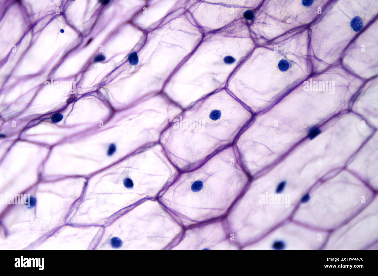
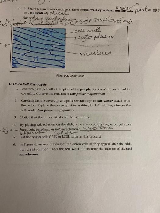
What color does the iodine stain the onion cell parts? Iodine- dark stain that colors starches in cells. In an onion cell, it will make the cell wall more visible. It provides some contrast for viewing under a microscope. Methylene Blue- a blue stain that will color blood, bacteria, acidic or protein rich cell structures like nucleus, ribosomes, and endoplasmic reticulum.

OBSERVING ONION PEEL EPIDERMAL CELLS UNDER MICROSCOPE | BEST DEMO | BIOLOGY
The Cell Structure of an Onion | Sciencing Onion cells are among the most common choices for cell studies in early biology classes. Easily obtained, inexpensive, they offer samples with no difficult technique required. The thin layer of skin found on the inside of an onion scale (one layer of onion) lifts off without effort and can be wet mounted on a slide with no need for extreme skill.
Onion Skin Cells - Investigation
DOC Plant and Animal Cells Microscope Lab - hillsboro.k12.oh.us Make a drawing of one onion cell, labeling all of its parts as you observe them. (At minimum you should observe the nucleus, cell wall, and cytoplasm.) Cheek cells 1. To view cheek cells, gently scrape the inside lining of your cheek with a toothpick. DO NOT GOUGE THE INSIDE OF YOUR CHEEK! (We will observe blood cells in a future lab!!) 2.
Solved I did a bio lab where I looked at purple onion skin ...
Solved 4. Using the low power objective, view several potato ...

3,009 Onion Cells Stock Photos, Pictures & Royalty-Free ...

Onion skin cells under the microscope | iPad Case & Skin
Solved] Fig. 2.7. Stained cells of fleshy onion (Allium cepa ...
Biology
Intro to cells (article) | Khan Academy
Mic-UK: The inner epidermis of the onion bulb's cataphylls ...

Sketch the onion peel cell as seen under the microscope ...

Staining of Onion Cell Nuclei | Microbehunter Microscopy

Tonicity-Onion cell lab | Miranda's AP Biology Blog
File:Onion cells under the light microscope.jpg - Wikimedia ...

How to Observe Onion Cells under a Microscope - Blog, She Wrote
STANDARD OPERATING PROCEDURE:
How is an onion's cell nucleus like? - Quora
Mic-UK: How many onion skins are there?
Post a Comment for "41 onion cells under microscope with labels"